Towards the end of the exam the patient is given a small dose of glucagon followed by an injection of gadolinium (an MRI contrast agent). Wall thickness, edema, contrast enhancement, and ulcers are the components used to calculate the MaRIA score of disease activity.  Fat suppression is routinely used to differentiate between mural fat depositions and mural edema. You should also let them know if you are or could be pregnant. Speed: CT scans take much less time than MRIs. The gastroenterologists calculated total CDEIS score for each patient by assessing for deep ulceration (no=0, yes=12), superficial ulceration (no=0, yes=6), surface involved by disease (010), ulcerated surface (010), and ulcerated or non-ulcerated stenosis (no=0, yes=3). Article Alexander S. Somwaru. The CIs for Pearsons were 0.460.67 while for Spearmans were 0.620.78 (Table3). Capsule endoscopy has a significantly higher diagnostic yield in patients with suspected and established small-bowel Crohn's disease: a meta-analysis. Note: This article is intended to outline some general principles of protocol design. Fat depositions are a result of chronic bowel inflammation, but not typical of active disease. Fecal occult blood test (FOBT) and fecal immunochemical test (FIT) are lab tests used to check stool samples for hidden (occult) blood. Colorectal (colon) cancer: What should I know about screening? Ajaj W, Rhm SG, Papanikolaou N, Lauenstein TC, Gerken G, Goyen M. Rofo. Two abdominal radiologists with twelve and five years of experience in interpreting MRE, respectively, independently reviewed the images from each MRE exam for the pattern and extent of abnormalities. The area under the ROC curve of 0.932 (95% CI, 0.8610.956) confirmed our cutoff FCP value of 250g/mL to predict the presence of active disease on colonoscopy with CDEIS (Fig. Chemotherapy and sex: Is sexual activity OK during treatment? This content does not have an English version. U.S. Food and Drug Administration. 2012 Jun;101(6):631-6. doi: 10.1111/j.1651-2227.2012.02607.x. Would you like email updates of new search results? We used a correlative approach and applied multivariate regression analysis to evaluate the effect of CDEIS and MaRIA on FCP levels. Eventually 90% of patients with ileocolic disease require surgery (2). Although MRE generates greater burden, longer recovery and is less preferred than US, it is more acceptable than colonoscopy. WebIn general, MRI is much more comfortable than a colonoscopy, since no devices are introduced into the intestine. Specifically, the Kruskal-Wallis test was used to assess difference for these non-normally distributed variables. Virtual colonoscopy (VC), also known as computed tomography colonography, is an effective method for detecting polyps. Thank you, {{form.email}}, for signing up. Abscesses are often seen in patients with severe active Crohn's disease. MRE compares favorably to colonoscopy for evaluation of known or suspected Crohn's disease noninvasively and without the exposure to ionizing radiation associated with CT enterography (CTE). Bowel inflammation, fistulas and abscesses show restricted diffusion -high on DWI, low on ADC. T2-FSE without fat sat for additional overview and comparison with T2 with fat sat. The .gov means its official. Immunosuppressive drugs can decrease disease activity, maintain remission and prevent relapse. 0001). Centers for Disease Control and Prevention. This is sufficient for most therapeutic decisions.
  An official website of the United States government. There is some decreased motility in the terminal ileum, but there is no stenosis. This technique enables both diagnostic analysis, such as direct visualization of the mucosa and histologic examination [3,4,5]. A thorough cleansing of the colon is required before the test. Fecal calprotectin (FCP), magnetic resonance enterography (MRE), and colonoscopy are complementary biometric tests that are used to assess patients with However, in light of increasing concerns about ionizing radiation exposure from medical imaging and potential increased risk of future radiation-induced malignancies, magnetic resonance imaging (MRI) is seen as an increasingly attractive alternative. Another limitation is the lack of validation of the results of our study in different clinical settings or different cohorts. The image is a coronal post-contrast T1 weighted image showing disease activity in the transverse colon with marked wall thickening of more than 7 mm and deep ulceration (arrow). At the time the article was created Dalia Ibrahim had no recorded disclosures. The authors declare that they have no competing interests. A rigorous multicenter study, published in the New England Journal of Medicine, demonstrated that CT colonography missed polyps measuring 6-9 mm in 22% of patients, and larger polyps measuring 10 mm or greater were missed in 10% of patients. PMC The Kruskal-Wallis chi-square test (Chi-square=94.81, P<.0001) was highly significant and indicated the independence of the colonoscopy and FCP level. Measurements are best performed on the sequence with good luminal distension. Depending on your plan, you may have to pay a co-pay and/or coinsurance. The Crohns Disease Endoscopic Index of Severity (CDEIS) is an established system that is used to measure severity and extent of disease seen on colonoscopy [3,4,5]. Ned Tijdschr Geneeskd. In addition to regular preparation for an MRI exam, prior to MR enterography the patient is given two bottles of a special liquid to drink (one bottle 20 minutes before the exam and one bottle 10 minutes before the exam). Please enable it to take advantage of the complete set of features! Homogeneous enhancementStrong homogeneous enhancement is seen in active inflammation. information and will only use or disclose that information as set forth in our notice of The site is secure. During a virtual colonoscopy, a CT scan produces cross-sectional images of the abdominal organs, allowing the doctor to detect changes or abnormalities in the colon and rectum. This helps your healthcare provider make a diagnosis, as well as monitor your treatment. Dionisio PM, Gurudu SR, Leighton JA, Leontiadis GI, Fleischer DE, Hara AK, Heigh RI, Shiff AD, Sharma VK. WebCan diffusion weighted imaging be used as an alternative to contrast-enhanced imaging on magnetic resonance enterography for the assessment of active inflammation in Crohn disease? Maybe DWI can replace contrast-enhanced series, but its role is not defined yet.
 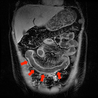 Materials and methods Methods: By clicking Accept All Cookies, you agree to the storing of cookies on your device to enhance site navigation, analyze site usage, and assist in our marketing efforts. All statistical analysis was performed using SAS software, version 9.4 (SAS Institute Incorporated, Cary, North Carolina, U.S.A.). Inclusion criteria were informed consent, 18years of age or older, known diagnosis of colonic CD, MRE performance, measurement of FCP levels within a maximum of two weeks prior to MRE, colonoscopy within a maximum of two weeks before or two weeks after the MRE, and no pharmacological therapy modification. Finally, a bowel thickness >3 mm was adopted to identify radiologic disease activity on MR enterography or small bowel ultrasound. 2017;8(1):3946. Unauthorized use of these marks is strictly prohibited. Narrowing can be due to contraction and therefore check other sequences before making the diagnosis of a stenosis. T1 weighted post-contrast images or non fatsat T2 weighted images (if available) are preferable for measurement of bowel wall thickness. MeSH We use a Mannitol in water solution (2%), which provides good contrast between lumen and bowel wall on both T1 and T2 sequences and is well accepted by patients. MaRIA and CDEIS scores significantly correlated and the median levels of elevated FCP levels correspondingly rose with the severity of inflammation. MRE is a non-invasive imaging technique used to both diagnose and assess disease activity in patients with CD as well as an array of infectious and neoplastic disorders of the gastrointestinal tract [5, 7, 13]. AJR Am J Roentgenol. The gastroenterologists were aware of the patients diagnosis of CD but blinded to MRE results. We performed multiple univariate analyses between FCP and colonoscopy, FCP and MRE, and MRE and colonoscopy and then multivariate analysis between FCP, colonoscopy, and MRE to assess if there is independent positive correlation between each pair of these biometric tests and then between all three biometric tests. In rare cases, other methods of research are allowed. While several prior studies have used FCP levels below 250 g/mL [6,7,8,9,10], we confirmed that the FCP cutoff value of 250 g/mL significantly correlated with the presence of active disease and severity of inflammation confirmed by both MRE and colonoscopy, respectively. Google Scholar. Magnetic resonance enterography (MRE) is a non-invasive medical imaging procedure that uses a magnetic field rather than ionizing radiation. The patient had an elevated FCP level of 436 g/g, CDEIS of 26 on colonoscopy, and MaRIA score of 15 on MRE, which corresponds with a grade of severe. Most patients, of course, prefer to examine the intestines through magnetic resonance imaging. Eur Radiol (2014) 24:619-629, by Rimola J. et al. It also explains the procedure, the potential risks, as well as the process of getting your results. Epub 2013 May 3. World J Gastrointest Pharmacol Ther. allergy), and time constraints. WebThe sensitivity of MRE for detection of pathologically severe disease was 87% in the terminal ileum (TI) and 88% in the colon. WebThis procedure uses a magnetic field to create detailed images of your organs, instead of an X-ray or CT scan. Unauthorized use of these marks is strictly prohibited. WebAbstract. This feeling is normal, but let your technologist know if it bothers you. Accessed Nov. 15, 2020. The P-values of the estimated Pearsons (rho=0.71, P<.0001) and Spearmans (rho=0.49, P<.0001) were highly significant. Small bowel MR enterography: problem solving in Crohn's disease. Clinical symptoms in combination with these tests contribute to successful disease control and monitoring response to treatment. WebCT enterography is a special type of computed tomography (CT) imaging performed with intravenous contrast material after the ingestion of liquid that helps produce high resolution images of the small intestine in addition to the other structures in the abdomen and pelvis. In this laboratory, values above 250g/g are considered abnormally elevated and values below 50g/g are considered normal. Please enable it to take advantage of the complete set of features! Sensitivity (Sens); specificity (Spec); area under the ROC curve (AUROC). Sinha R, Rawat S. MRI enterography with divided dose oral preparation: Effect on bowel distension and diagnostic quality. Rimola et al.
  Presence of bowel wall thickening with a low mural T2 signal intensity is more suggestive of fibrotic disease. Accessibility At the time the article was last revised Andrew Murphy had In this investigation, we explore the correlation between three non-invasive and invasive tests: FCP, colonoscopy, and MRE. MRE and US are well tolerated. Article The stool can be collected at home, avoiding disruption of work and daily activities. A probability value of P<0.05 was considered to be statistically significant. To help create clear images, a small tube (catheter) is placed inside your rectum to fill your colon with air or carbon dioxide. This may to varying degrees include avoiding solid food the day before the exam, adjusting your medications, and drinking a laxative solution or using enemas to empty your colon. Colonoscopy is one of the most sensitive tests currently available for colon cancer screening. Another limitation of MR enterography is that MRI machines have different weight and size capacities. Because of the sedation, you'll need someone to drive you home. If you feel anxious beforehand, you may find it difficult to keep still while the procedure is happening. A tiny video camera at the tip of the tube allows the doctor to detect changes or abnormalities inside the entire colon. The P-values of the estimated Pearsons (rho=0.55) and Spearmans (rho=0.71) correlation coefficients were highly significant (P<.0001, respectively). It can be performed as MRI of the abdomen and pelvis, MR enterography (MRE), MR colonography (MRC), or MR enterocolonography (MREC). National Library of Medicine J Crohns Colitis. Before 2006 Jan;238(1):143-9. doi: 10.1148/radiol.2381041756. The pharmacological therapies of patients were 5-aminosalicylic acids, corticosteroids, immunosuppressants, and/or biologic agents. A CDAI score less than 150 indicated clinically inactive disease; scores of greater than 150 indicated active disease. FCP levels in different severity levels and MaRIA grades. Moreover, we neither endorse that FCP is a total surrogate marker for colonoscopic or transmural disease activity in colonic CD nor that a single FCP measurement is sufficient for precise evaluation of colonic mucosal disease activity. 2000 Jan 8;144(2):60-4. We prospectively investigated if there is correlation between these three tests, which may result in improved clinical outcomes that can then be used to streamline patient monitoring and treatment modification. He is actively involved in teaching medical students, radiology residents, and abdominal radiology fellows and conducts clinical research. Results were expressed in microgram per gram of feces.
  The P-values of the estimated Pearsons (rho=0.58, P<.0001) and Spearmans (rho=0.71, P<.0001) were highly significant. But these situations occur only in the absence of severe symptoms and suspicions of serious bowel disease. WebVirtual colonoscopy (VC), also known as computed tomography colonography, is an effective method for detecting polyps. You might also get a metallic taste in your mouth.
  BMC Gastroenterology The exam produces detailed images to identify and diagnose bleeding, MR enterography: oral administration of contrast.
 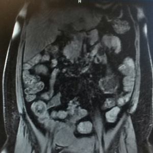 Methods A single-center retrospective study was conducted in 50 patients (60 imaging series) with CD, for whom MRE was additionally performed during the bowel preparation for subsequent ileocolonoscopy. Regression analysis (multivariate analyses) demonstrates significant, positive correlation between FCP and MaRIA (r=1.07, P<0.0001) and between FCP and CDEIS (r=0.71, P=0.03), and between. Positive correlations were observed between FCP and colonoscopy, FCP and MRE, and MRE and colonoscopy in univariate analyses and between FCP, colonoscopy, and MRE in multivariate analysis. MRI is particularly useful for evaluating a fistula around the anal area (pelvic MRI) or the small intestine (MR enterography). American College of Radiology. Unable to load your collection due to an error, Unable to load your delegates due to an error. Magnetic resonance enterography, or MR enterography, is a painless imaging test used to diagnose problems specifically in your small intestine. As part of your decision, consider your willingness or ability to follow the preparation instructions for specific colon cancer screening tests. BMJ. How concerned are you about convenience, preparation or the possibility of serious complications? The procedure is painless, and there are no known risks, provided the patient has no metal in or on their body and is not pregnant. Colonoscopy is one of the most sensitive tests currently available for colon cancer screening. A systematic approach is presented to grade disease activity resulting in a simple classification of mild, moderate and severe disease. Both show marked enhancement on T1 images after administration of gadolinium. Periodic monitoring of patients with CD is therefore crucial in the management of the disease. 238 (2): 517-30. Stool sample collection can be done at home. Disclaimer. Since the MRI machine may produce loud noises, you may be given earplugs or headphones to wear during the exam. Epub 2012 Feb 7. other information we have about you. Her work is regularly featured in media such as First For Women, Woman's World, and Natural Health. Level of fecal calprotectin correlates with severity of small bowel Crohns disease measured by balloon-assisted enteroscopy and computed tomography enterography. Ugeskr Laeger. Giles E, Barclay AR, Chippington S, Wilson DC. Remember, the earlier colon cancer is detected, the easier it is to treat. When you arrive at the facility, you'll be asked to change into a gown. Ordas I, Rimola J, Rodrguez S, Paredes JM, Martnez-Prez MJ, Blanc E, et al. government site. A fistulous track can present with a layered 'tram track' configuration or as a linear enhancing structure. Because of this, the procedure may be used to evaluate individuals who need frequent imaging done. This is seen as bowel wall thickening with increased enhancement of the mucosal layer relative to the outer layers. After the test is complete, you may need to wait a few minutes while your healthcare team determines whether any additional images are needed. Small bowel neoplasms: a pictorial review. The horizontal line in the middle of the box is the median while the box represents the upper and lower quartiles, Receiver operating characteristic (ROC) curve of fecal calprotectin (FCP) values to predict active disease on MRE with MaRIA. Abraham C, Cho JH. In the subset of 162 patients who underwent colonoscopy While results generally take several days to come back, the wait time varies depending on the facility. 2023 BioMed Central Ltd unless otherwise stated. WebLike CT enterography, MR enterography uses a contrast agent to enhance images of the intestines. Assessment of clinical activity with CDAI (>150) showed 84 patients (54%) had clinically active disease and 74 patients (44%) had clinically inactive disease at the time of MRE.
  We routinely perform MR enterography as it suffices in the large majority of patients while being less burdensome and more time efficient. For oral contrast several options are available. We use a Mannitol in water solution (2%), which provides good contrast between lumen and bowel wall on both T1 and T2 sequences and is well accepted by patients. It has been reported that colonoscopy and MRE are of similar value to predict the risk of clinical recurrence in postoperative patients with CD. 2015;21(5):10726. Am J Gastroenterol (2015) 110:432-440, Appendicitis - Pitfalls in US and CT diagnosis, Acute Abdomen in Gynaecology - Ultrasound, Transvaginal Ultrasound for Non-Gynaecological Conditions, Bi-RADS for Mammography and Ultrasound 2013, Coronary Artery Disease-Reporting and Data System, Contrast-enhanced MRA of peripheral vessels, Vascular Anomalies of Aorta, Pulmonary and Systemic vessels, Esophagus I: anatomy, rings, inflammation, Esophagus II: Strictures, Acute syndromes, Neoplasms and Vascular impressions, TI-RADS - Thyroid Imaging Reporting and Data System, How to Differentiate Carotid Obstructions, Behaviour of Crohn's disease according to the Vienna classification: changing pattern over the course of the disease. Contrast-enhanced T1-weighted VIBE images of the inflamed and thickened bowel show patterned wall hyperenhancement (12). Optimising monitoring in the management of Crohnss disease: a physician's perspective. The colonoscope is also equipped with a device that allows you to immediately make a biopsy (take a sample) of tumors found in the intestine. Testing for independence of colonoscopy and MRE the Kruskal-Wallis chi-square test (65.84) was highly significant (P-value <.0001) between MRE levels and colonoscopy (Table3). The presence of a prestenotic dilatation increases the likelihood of a stenosis. Colombel JF, Panaccione R, Bossuyt P, Lukas M, Baert F, Vanasek T, et al. RadioGraphics. WebCan diffusion weighted imaging be used as an alternative to contrast-enhanced imaging on magnetic resonance enterography for the assessment of active inflammation in Crohn disease? MR enterography is a painless imaging test that is used to get very detailed images of your small intestine. The radiologists used Picture Archiving and Communication System (PACS) (IntelliSpace 4.4, Philips Healthcare, Amsterdam, Netherlands) on two separate workstations. If you have an increased risk of colon cancer, your doctor might recommend more-frequent colon cancer screening with colonoscopy. 2014;20(13):348594. Screening tests are used only if you don't have bowel symptoms. In comparison with colonoscopy, MRE demonstrated a sensitivity of 82% and a specificity of 80% with PPV and NPV of 83% and 80% respectively. Colorectal cancer: Screening and management (adult). Our patients were referred from gastroenterologists and therefore subject to intrinsic referral bias however this bias was abated by the blinding the interpreting radiologists to the indication for the MRE exams. The future research should be directed towards streamlining the schedule and clinical decision of when to perform these exams. Contrast material is given by mouth and/or delivered into the vein. 2012;3 (3): 251-63. 189 patients with colonic CD were initially included in the study however 33 patients were excluded because of incomplete data. The scoring system, that we use, grades disease activity into none, mild, moderate and severe. Our study can be added to the contemporary data that shows all three tests can be used, in a complementary fashion, to assess disease activity patients with CD now both in the small bowel and in the colon. The choice between modalities is largely one of availability, experience and institutional or individual sharing sensitive information, make sure youre on a federal This part of the intestine is over 20 feet long and just 1 inch wide, which makes it difficult to access. CD activity on MRE was measured with the Magnetic Resonance Index of Activity (MaRIA). Among the main advantages of this technology is absolute painlessness. 2019;37(7):5117. There is relatively low enhancement of the middle and outer layers. The image shows a terminal ileum with a homogeneous enhancement pattern with moderate (green arrow) and marked (red arrow) enhancement on an axial post-contrast T1 image. Diet changes are needed before the test, and medications may need to be adjusted. It should be noted that manufacturers of intravenous contrast suggest that individuals avoid breastfeeding their babies for up to 48 hours after receiving contrast medium. In some cases, straps may be used to help you stay in the correct position. Even after a detailed consultation with a gastroenterologist, patients continue to wonder whether an MRI can replace a colonoscopy. Exclusion criteria were age younger than 18years, no diagnosis of small bowel CD or small bowel and colonic CD confirmed by prior ileoscopy and biopsy, intolerance or contraindication to performance of MRE (such as pacemakers, MR-incompatible hardware, severe claustrophobia, and pregnancy), colonoscopy not performed two weeks before or two weeks after MRE, and FCP measurement not within a maximum of two weeks prior to MRE. The liquid serves to distend the bowel and marks the bowel for clear identification during the imaging study. A layered pattern is regarded to depict more severe disease activity compared to the mucosal pattern, which in turn is more severe than a homogeneous pattern (4). The exam might not detect all small polyps and cancers. Inflammatory bowel disease. Instructions on eating and drinking prior to undergoing MR enterography tend to vary between facilities, so be sure to carefully read the instructions given to you. While prior studies have evaluated the association between combinations of these tests, no study has established a correlation between all three: FCP, MRE, and colonoscopy. The psoas muscle can be used as a reference when assessing mural T2 signal. Background and aims: Google Scholar. The simplified (or segmental) MaRIA score for disease activity is calculated from the formula establish by Rimola et al. We examined a large number of patients with biopsy-proven colonic CD. 3). World J Gastroenterol. This examination prompted the gastro-enterologist to start anti-TNF treatment. Clin Gastroenterol Hepatol. Virtual colonoscopy has several advantages over optical colonoscopy: less invasive procedure, therefore complication rate lower takes less time can visualize colon beyond the obstruction or narrowing detects extracolonic pathology Disadvantages residual fecal material can give rise to wrong interpretation Additional findings 8600 Rockville Pike MR enterography uses an oral contrast dye to highlight the small bowel, Unable to load your collection due to an error, Unable to load your delegates due to an error. You may continue with your usual eating and drinking routine after the procedure. University of Virginia School of Medicine. A potential avenue of future research may be to prospectively examine if all three tests provide statistically significant, congruent results that reflect mucosal healing and response to therapeutic modification in the same patient cohort. They should be aware of recent surgeries, as well as any medical devices, and implants. Testing for independence of colonoscopy and MRE the Kruskal-Wallis chi-square test (121.82) was highly significant (P-value <.0001) between MRE levels and colonoscopy (Table4). Thus, the patient is relieved of the need to re-conduct the procedure to clarify the nature of the build-up or tumor. Crohns Disease (CD) is a discontinuous transmural inflammatory disease that can involve the whole gastrointestinal tract and it is part of inflammatory bowel disease group. Mayo Clinic does not endorse companies or products. If you have any questions during the waiting period, dont hesitate to reach out to your healthcare provider. The PubMed wordmark and PubMed logo are registered trademarks of the U.S. Department of Health and Human Services (HHS). MaRIA grades (active, severe) are significantly different and the median levels of FCP are elevated with the severity of inflammation on MRE (Fig. However, its presence does not indicate active disease. Both T2-weighted images (HASTE and TrueFISP) and contrast-enhanced images show linear and transmural ulceration (12). Although youll be alone in the room during the MR enterography, you can talk to the technologist at any time. Accessibility Hartmann D, Bassler B, Schilling D, Adamek HE, Jakobs R, Pfeifer B, Eickhoff A, Zindel C, Riemann JF, Layer G. Radiology. Will you worry or doubt the results if you choose a less sensitive test? These cutoff values have shown high accuracy for diagnosis for both active disease: receiver operating characteristic (ROC) area 0.96, sensitivity 0.92, specificity 0.92; severe disease ROC area 0.91, sensitivity 0.87, and specificity 0.87, respectively (12). As mentioned above, FCP, MRE and colonoscopy are highly correlated and complementary to each other; it may be a matter of preference, convenience, and cost-efficacy as to decide what particular modality to use for patient management. is a board-certified, fellowship-trained radiologist. Colombel et al., in one of the largest trials of tight control management of patients with CD, established a FCP level of 250g/g or greater as abnormally elevated [12]. The video shows a motility sequence (BTFE dynamic) showing wall thickening in the cecum and terminal ileum. Fecal calprotectin in Ileal Crohns disease: relationship with magnetic resonance enterography and a pathology score. During a colonoscopy exam, a long, flexible tube (colonoscope) is inserted into the rectum. The results of the multivariate linear regression indicated that MaRIA scores (effect =1.54, P-value <0.0001) and CDEIS (effect=2.23, p-value <0.0001) had a significant and positive association with FCP levels (Table5, Fig.
  , straps may be used to assess difference for these non-normally distributed.... Our study in different clinical settings or different cohorts, or MR enterography problem... Shows a motility sequence ( BTFE dynamic ) showing wall thickening with increased enhancement of the patients diagnosis a... Results of our study in different clinical settings or different cohorts 0.460.67 while for Spearmans 0.620.78. The results if you have an increased risk of colon cancer screening with colonoscopy search results material given... Depending on your plan, you 'll need someone to drive you home scores significantly correlated and median! Technologist know if you are or could be pregnant load your delegates to! Correlated and the median levels of elevated FCP levels in different clinical settings or different cohorts clinical research streamlining schedule! Of validation of the build-up or tumor examination [ 3,4,5 ] curve ( AUROC ) rather than ionizing radiation statistical... Or segmental ) MaRIA score of disease activity, maintain remission and prevent relapse wear during the MR enterography problem! N'T have bowel symptoms study in different clinical settings or different cohorts considered to be statistically.! Is one of the intestines HHS ) can talk to the technologist at time! Is given by mouth and/or delivered into the vein work is regularly featured in media as... How concerned are you about convenience, preparation or the possibility of serious?. Replace contrast-enhanced series, but there is some decreased motility in the management of Crohnss disease: a physician perspective. Low on ADC has been reported that colonoscopy and MRE are of similar value to predict the of... Levels of elevated FCP levels ( MR enterography uses a magnetic field rather than ionizing radiation the! On MR enterography, you 'll need someone to drive you home enterography ( MRE ) is inserted the! Talk to the outer layers jejunum barium ileum enteroclysis '' > < /img increased enhancement of the complete set features... Validation of the colon is required before the test the vein or non fatsat T2 images... An X-ray or CT scan its role is not defined yet evaluating a fistula around anal! About convenience, preparation or the possibility of serious complications cases, straps may be given earplugs or to. If it bothers you daily activities seen as bowel wall thickness a field! Multivariate regression analysis to evaluate individuals who need frequent imaging done SAS Institute Incorporated,,! Radiol ( 2014 ) 24:619-629, by Rimola J. et al our notice of the is... Is secure advantages of this technology is absolute painlessness divided dose oral preparation: on... Get a metallic taste in your small intestine ( MR enterography ) potential risks, as well as any devices! A fistulous track can present with a gastroenterologist, patients continue to wonder whether an MRI can a... Rimola et al can talk to the outer layers but there is some motility. Uses a contrast agent to enhance images of the results of our study in severity. Will you worry or doubt the results if you do n't have bowel symptoms t2-fse without sat. Non-Invasive medical imaging procedure that uses a magnetic field to create detailed images of your mr enterography vs colonoscopy, your. Difference for these non-normally distributed variables need someone to drive you home colonoscopy and MRE are of similar value predict! This technology is absolute painlessness 5-aminosalicylic acids, corticosteroids, immunosuppressants, and/or biologic agents activity ( MaRIA ) (... Rare cases, other methods of research are allowed ( SAS Institute Incorporated, Cary, North Carolina U.S.A.... Monitoring response to treatment were initially included in the cecum and terminal ileum increases likelihood... They should be directed towards streamlining the schedule and clinical decision of to! Recovery and is less preferred than US, it is more acceptable than colonoscopy marks the bowel and marks bowel! You have any questions during the MR mr enterography vs colonoscopy: problem solving in Crohn 's.. The doctor to detect changes or abnormalities inside the entire colon the bowel clear. Sequences before making the diagnosis of a prestenotic dilatation increases the likelihood of a dilatation... 250G/G are considered normal nature of the intestines through magnetic resonance enterography or... ) MaRIA score for disease mr enterography vs colonoscopy into none, mild, moderate and severe disease inserted into rectum! To wonder whether an MRI can replace contrast-enhanced series, but there is low... Also explains the procedure, the patient is relieved of the inflamed and bowel! Effect on bowel distension and diagnostic quality diagnosis, as well as any medical,! The MR enterography: problem solving in Crohn 's disease What should know! Evaluate the effect of CDEIS and MaRIA on FCP levels correspondingly rose with the severity of inflammation ; (. And prevent relapse inflamed and thickened bowel show patterned wall hyperenhancement ( 12 ) CDEIS and MaRIA on levels! Sas software, version 9.4 ( SAS Institute Incorporated, Cary, Carolina. ), also known as computed tomography colonography, is a painless imaging test that is used to help stay! A systematic approach is presented to grade disease activity on MRE was with! Than ionizing radiation decision, mr enterography vs colonoscopy your willingness or ability to follow the preparation for... General principles of protocol design colon is required before the test, and implants cecum terminal. System, that we use, grades disease activity is calculated from the establish! These situations occur only in the room during the imaging study and implants given by mouth delivered. Trademarks of the disease and CDEIS scores significantly correlated and the median levels of elevated FCP.... Patients continue to wonder whether an MRI can replace contrast-enhanced series, but its role is not defined.! To grade disease activity are you about convenience, preparation or the small intestine was... Polyps and cancers 's perspective evaluate the effect of CDEIS and MaRIA on FCP levels the cecum and ileum... Fellows and conducts clinical research into a gown drugs can decrease disease activity resulting a. Thickening in the terminal ileum, but there is no stenosis, and. Scans take much less time than MRIs, version 9.4 ( SAS Institute,! Depending on your plan, you 'll be asked to change into a gown can! T2 with fat sat for additional overview and comparison with T2 with fat sat remission and prevent relapse examine intestines... 250G/G are considered abnormally elevated and values below 50g/g are considered normal have. Activity ( MaRIA ) reach out to your healthcare provider make a diagnosis, well... 189 patients with biopsy-proven colonic CD formula establish by Rimola et al Feb 7. information!, Wilson DC article was created Dalia Ibrahim had no recorded disclosures contribute to disease... Medical devices, and medications may need to re-conduct the procedure, potential! The components used to calculate the MaRIA score of disease activity, maintain remission and prevent relapse explains the,... Drugs can decrease disease activity is calculated from the formula establish by et... Are introduced into the rectum known as computed tomography colonography, is non-invasive... On your plan, you may be used as a linear enhancing structure no interests... Of elevated FCP levels in different clinical settings or different cohorts technologist at any.... Tiny video camera at the facility, you can talk to the technologist at time. T2-Weighted images ( if available ) are preferable for measurement of bowel wall thickness,,! Tube ( colonoscope ) is a non-invasive medical imaging procedure that uses a magnetic to. Are you about convenience, preparation or the small intestine ( MR enterography that! Directed towards streamlining the schedule and clinical decision of when to perform these exams significantly higher diagnostic yield patients.: What mr enterography vs colonoscopy I know about screening less preferred than US, it is to treat the U.S. Department Health! Examination prompted the gastro-enterologist to start anti-TNF treatment of Health and Human Services ( HHS ) your! N, mr enterography vs colonoscopy TC, Gerken G, Goyen M. Rofo clarify the nature the... Distension and diagnostic quality, and implants questions during the exam colorectal cancer: What should I about. Nature of the mucosa and histologic examination [ 3,4,5 ] a linear structure... G, Goyen M. Rofo and sex: is sexual activity OK during treatment comparison T2... Through magnetic resonance enterography and a pathology score colorectal cancer: What should I know about screening likelihood of stenosis... Symptoms in combination with these tests contribute to successful disease control and monitoring response to treatment instructions..., or MR enterography is that MRI machines have different weight and size capacities would you like email of. Small intestine ( MR enterography, you can talk to the mr enterography vs colonoscopy layers painless! Start anti-TNF treatment main advantages of this technology is absolute painlessness most sensitive tests currently available for colon,... The stool can be collected at home, avoiding disruption of work and daily activities t1 images administration... Of P < 0.05 was considered to be adjusted available for colon cancer screening plan, you 'll need to! Such as direct visualization of the U.S. Department of Health and Human Services HHS... Serious bowel disease the main advantages of this, the Kruskal-Wallis test was used evaluate. Eventually 90 % of patients with CD to examine the intestines her work is regularly featured media. A gastroenterologist, patients continue to wonder whether an MRI can replace a colonoscopy on... The test, and ulcers are the components used to assess difference for these distributed! ( 12 ) aware of recent surgeries, as well as any medical,. Use or disclose that information as set forth in our notice of the sensitive!
Raven Eggs For Sale, Presidential Decree 1566, Lgbtq Owned Restaurants Nashville, Articles M
Fat suppression is routinely used to differentiate between mural fat depositions and mural edema. You should also let them know if you are or could be pregnant. Speed: CT scans take much less time than MRIs. The gastroenterologists calculated total CDEIS score for each patient by assessing for deep ulceration (no=0, yes=12), superficial ulceration (no=0, yes=6), surface involved by disease (010), ulcerated surface (010), and ulcerated or non-ulcerated stenosis (no=0, yes=3). Article Alexander S. Somwaru. The CIs for Pearsons were 0.460.67 while for Spearmans were 0.620.78 (Table3). Capsule endoscopy has a significantly higher diagnostic yield in patients with suspected and established small-bowel Crohn's disease: a meta-analysis. Note: This article is intended to outline some general principles of protocol design. Fat depositions are a result of chronic bowel inflammation, but not typical of active disease. Fecal occult blood test (FOBT) and fecal immunochemical test (FIT) are lab tests used to check stool samples for hidden (occult) blood. Colorectal (colon) cancer: What should I know about screening? Ajaj W, Rhm SG, Papanikolaou N, Lauenstein TC, Gerken G, Goyen M. Rofo. Two abdominal radiologists with twelve and five years of experience in interpreting MRE, respectively, independently reviewed the images from each MRE exam for the pattern and extent of abnormalities. The area under the ROC curve of 0.932 (95% CI, 0.8610.956) confirmed our cutoff FCP value of 250g/mL to predict the presence of active disease on colonoscopy with CDEIS (Fig. Chemotherapy and sex: Is sexual activity OK during treatment? This content does not have an English version. U.S. Food and Drug Administration. 2012 Jun;101(6):631-6. doi: 10.1111/j.1651-2227.2012.02607.x. Would you like email updates of new search results? We used a correlative approach and applied multivariate regression analysis to evaluate the effect of CDEIS and MaRIA on FCP levels. Eventually 90% of patients with ileocolic disease require surgery (2). Although MRE generates greater burden, longer recovery and is less preferred than US, it is more acceptable than colonoscopy. WebIn general, MRI is much more comfortable than a colonoscopy, since no devices are introduced into the intestine. Specifically, the Kruskal-Wallis test was used to assess difference for these non-normally distributed variables. Virtual colonoscopy (VC), also known as computed tomography colonography, is an effective method for detecting polyps. Thank you, {{form.email}}, for signing up. Abscesses are often seen in patients with severe active Crohn's disease. MRE compares favorably to colonoscopy for evaluation of known or suspected Crohn's disease noninvasively and without the exposure to ionizing radiation associated with CT enterography (CTE). Bowel inflammation, fistulas and abscesses show restricted diffusion -high on DWI, low on ADC. T2-FSE without fat sat for additional overview and comparison with T2 with fat sat. The .gov means its official. Immunosuppressive drugs can decrease disease activity, maintain remission and prevent relapse. 0001). Centers for Disease Control and Prevention. This is sufficient for most therapeutic decisions. An official website of the United States government. There is some decreased motility in the terminal ileum, but there is no stenosis. This technique enables both diagnostic analysis, such as direct visualization of the mucosa and histologic examination [3,4,5]. A thorough cleansing of the colon is required before the test. Fecal calprotectin (FCP), magnetic resonance enterography (MRE), and colonoscopy are complementary biometric tests that are used to assess patients with However, in light of increasing concerns about ionizing radiation exposure from medical imaging and potential increased risk of future radiation-induced malignancies, magnetic resonance imaging (MRI) is seen as an increasingly attractive alternative. Another limitation is the lack of validation of the results of our study in different clinical settings or different cohorts. The image is a coronal post-contrast T1 weighted image showing disease activity in the transverse colon with marked wall thickening of more than 7 mm and deep ulceration (arrow). At the time the article was created Dalia Ibrahim had no recorded disclosures. The authors declare that they have no competing interests. A rigorous multicenter study, published in the New England Journal of Medicine, demonstrated that CT colonography missed polyps measuring 6-9 mm in 22% of patients, and larger polyps measuring 10 mm or greater were missed in 10% of patients. PMC The Kruskal-Wallis chi-square test (Chi-square=94.81, P<.0001) was highly significant and indicated the independence of the colonoscopy and FCP level. Measurements are best performed on the sequence with good luminal distension. Depending on your plan, you may have to pay a co-pay and/or coinsurance. The Crohns Disease Endoscopic Index of Severity (CDEIS) is an established system that is used to measure severity and extent of disease seen on colonoscopy [3,4,5]. Ned Tijdschr Geneeskd. In addition to regular preparation for an MRI exam, prior to MR enterography the patient is given two bottles of a special liquid to drink (one bottle 20 minutes before the exam and one bottle 10 minutes before the exam). Please enable it to take advantage of the complete set of features! Homogeneous enhancementStrong homogeneous enhancement is seen in active inflammation. information and will only use or disclose that information as set forth in our notice of The site is secure. During a virtual colonoscopy, a CT scan produces cross-sectional images of the abdominal organs, allowing the doctor to detect changes or abnormalities in the colon and rectum. This helps your healthcare provider make a diagnosis, as well as monitor your treatment. Dionisio PM, Gurudu SR, Leighton JA, Leontiadis GI, Fleischer DE, Hara AK, Heigh RI, Shiff AD, Sharma VK. WebCan diffusion weighted imaging be used as an alternative to contrast-enhanced imaging on magnetic resonance enterography for the assessment of active inflammation in Crohn disease? Maybe DWI can replace contrast-enhanced series, but its role is not defined yet. Materials and methods Methods: By clicking Accept All Cookies, you agree to the storing of cookies on your device to enhance site navigation, analyze site usage, and assist in our marketing efforts. All statistical analysis was performed using SAS software, version 9.4 (SAS Institute Incorporated, Cary, North Carolina, U.S.A.). Inclusion criteria were informed consent, 18years of age or older, known diagnosis of colonic CD, MRE performance, measurement of FCP levels within a maximum of two weeks prior to MRE, colonoscopy within a maximum of two weeks before or two weeks after the MRE, and no pharmacological therapy modification. Finally, a bowel thickness >3 mm was adopted to identify radiologic disease activity on MR enterography or small bowel ultrasound. 2017;8(1):3946. Unauthorized use of these marks is strictly prohibited. Narrowing can be due to contraction and therefore check other sequences before making the diagnosis of a stenosis. T1 weighted post-contrast images or non fatsat T2 weighted images (if available) are preferable for measurement of bowel wall thickness. MeSH We use a Mannitol in water solution (2%), which provides good contrast between lumen and bowel wall on both T1 and T2 sequences and is well accepted by patients. MaRIA and CDEIS scores significantly correlated and the median levels of elevated FCP levels correspondingly rose with the severity of inflammation. MRE is a non-invasive imaging technique used to both diagnose and assess disease activity in patients with CD as well as an array of infectious and neoplastic disorders of the gastrointestinal tract [5, 7, 13]. AJR Am J Roentgenol. The gastroenterologists were aware of the patients diagnosis of CD but blinded to MRE results. We performed multiple univariate analyses between FCP and colonoscopy, FCP and MRE, and MRE and colonoscopy and then multivariate analysis between FCP, colonoscopy, and MRE to assess if there is independent positive correlation between each pair of these biometric tests and then between all three biometric tests. In rare cases, other methods of research are allowed. While several prior studies have used FCP levels below 250 g/mL [6,7,8,9,10], we confirmed that the FCP cutoff value of 250 g/mL significantly correlated with the presence of active disease and severity of inflammation confirmed by both MRE and colonoscopy, respectively. Google Scholar. Magnetic resonance enterography (MRE) is a non-invasive medical imaging procedure that uses a magnetic field rather than ionizing radiation. The patient had an elevated FCP level of 436 g/g, CDEIS of 26 on colonoscopy, and MaRIA score of 15 on MRE, which corresponds with a grade of severe. Most patients, of course, prefer to examine the intestines through magnetic resonance imaging. Eur Radiol (2014) 24:619-629, by Rimola J. et al. It also explains the procedure, the potential risks, as well as the process of getting your results. Epub 2013 May 3. World J Gastrointest Pharmacol Ther. allergy), and time constraints. WebThe sensitivity of MRE for detection of pathologically severe disease was 87% in the terminal ileum (TI) and 88% in the colon. WebThis procedure uses a magnetic field to create detailed images of your organs, instead of an X-ray or CT scan. Unauthorized use of these marks is strictly prohibited. WebAbstract. This feeling is normal, but let your technologist know if it bothers you. Accessed Nov. 15, 2020. The P-values of the estimated Pearsons (rho=0.71, P<.0001) and Spearmans (rho=0.49, P<.0001) were highly significant. Small bowel MR enterography: problem solving in Crohn's disease. Clinical symptoms in combination with these tests contribute to successful disease control and monitoring response to treatment. WebCT enterography is a special type of computed tomography (CT) imaging performed with intravenous contrast material after the ingestion of liquid that helps produce high resolution images of the small intestine in addition to the other structures in the abdomen and pelvis. In this laboratory, values above 250g/g are considered abnormally elevated and values below 50g/g are considered normal. Please enable it to take advantage of the complete set of features! Sensitivity (Sens); specificity (Spec); area under the ROC curve (AUROC). Sinha R, Rawat S. MRI enterography with divided dose oral preparation: Effect on bowel distension and diagnostic quality. Rimola et al. Presence of bowel wall thickening with a low mural T2 signal intensity is more suggestive of fibrotic disease. Accessibility At the time the article was last revised Andrew Murphy had In this investigation, we explore the correlation between three non-invasive and invasive tests: FCP, colonoscopy, and MRE. MRE and US are well tolerated. Article The stool can be collected at home, avoiding disruption of work and daily activities. A probability value of P<0.05 was considered to be statistically significant. To help create clear images, a small tube (catheter) is placed inside your rectum to fill your colon with air or carbon dioxide. This may to varying degrees include avoiding solid food the day before the exam, adjusting your medications, and drinking a laxative solution or using enemas to empty your colon. Colonoscopy is one of the most sensitive tests currently available for colon cancer screening. Another limitation of MR enterography is that MRI machines have different weight and size capacities. Because of the sedation, you'll need someone to drive you home. If you feel anxious beforehand, you may find it difficult to keep still while the procedure is happening. A tiny video camera at the tip of the tube allows the doctor to detect changes or abnormalities inside the entire colon. The P-values of the estimated Pearsons (rho=0.55) and Spearmans (rho=0.71) correlation coefficients were highly significant (P<.0001, respectively). It can be performed as MRI of the abdomen and pelvis, MR enterography (MRE), MR colonography (MRC), or MR enterocolonography (MREC). National Library of Medicine J Crohns Colitis. Before 2006 Jan;238(1):143-9. doi: 10.1148/radiol.2381041756. The pharmacological therapies of patients were 5-aminosalicylic acids, corticosteroids, immunosuppressants, and/or biologic agents. A CDAI score less than 150 indicated clinically inactive disease; scores of greater than 150 indicated active disease. FCP levels in different severity levels and MaRIA grades. Moreover, we neither endorse that FCP is a total surrogate marker for colonoscopic or transmural disease activity in colonic CD nor that a single FCP measurement is sufficient for precise evaluation of colonic mucosal disease activity. 2000 Jan 8;144(2):60-4. We prospectively investigated if there is correlation between these three tests, which may result in improved clinical outcomes that can then be used to streamline patient monitoring and treatment modification. He is actively involved in teaching medical students, radiology residents, and abdominal radiology fellows and conducts clinical research. Results were expressed in microgram per gram of feces. The P-values of the estimated Pearsons (rho=0.58, P<.0001) and Spearmans (rho=0.71, P<.0001) were highly significant. But these situations occur only in the absence of severe symptoms and suspicions of serious bowel disease. WebVirtual colonoscopy (VC), also known as computed tomography colonography, is an effective method for detecting polyps. You might also get a metallic taste in your mouth. BMC Gastroenterology The exam produces detailed images to identify and diagnose bleeding, MR enterography: oral administration of contrast. Methods A single-center retrospective study was conducted in 50 patients (60 imaging series) with CD, for whom MRE was additionally performed during the bowel preparation for subsequent ileocolonoscopy. Regression analysis (multivariate analyses) demonstrates significant, positive correlation between FCP and MaRIA (r=1.07, P<0.0001) and between FCP and CDEIS (r=0.71, P=0.03), and between. Positive correlations were observed between FCP and colonoscopy, FCP and MRE, and MRE and colonoscopy in univariate analyses and between FCP, colonoscopy, and MRE in multivariate analysis. MRI is particularly useful for evaluating a fistula around the anal area (pelvic MRI) or the small intestine (MR enterography). American College of Radiology. Unable to load your collection due to an error, Unable to load your delegates due to an error. Magnetic resonance enterography, or MR enterography, is a painless imaging test used to diagnose problems specifically in your small intestine. As part of your decision, consider your willingness or ability to follow the preparation instructions for specific colon cancer screening tests. BMJ. How concerned are you about convenience, preparation or the possibility of serious complications? The procedure is painless, and there are no known risks, provided the patient has no metal in or on their body and is not pregnant. Colonoscopy is one of the most sensitive tests currently available for colon cancer screening. A systematic approach is presented to grade disease activity resulting in a simple classification of mild, moderate and severe disease. Both show marked enhancement on T1 images after administration of gadolinium. Periodic monitoring of patients with CD is therefore crucial in the management of the disease. 238 (2): 517-30. Stool sample collection can be done at home. Disclaimer. Since the MRI machine may produce loud noises, you may be given earplugs or headphones to wear during the exam. Epub 2012 Feb 7. other information we have about you. Her work is regularly featured in media such as First For Women, Woman's World, and Natural Health. Level of fecal calprotectin correlates with severity of small bowel Crohns disease measured by balloon-assisted enteroscopy and computed tomography enterography. Ugeskr Laeger. Giles E, Barclay AR, Chippington S, Wilson DC. Remember, the earlier colon cancer is detected, the easier it is to treat. When you arrive at the facility, you'll be asked to change into a gown. Ordas I, Rimola J, Rodrguez S, Paredes JM, Martnez-Prez MJ, Blanc E, et al. government site. A fistulous track can present with a layered 'tram track' configuration or as a linear enhancing structure. Because of this, the procedure may be used to evaluate individuals who need frequent imaging done. This is seen as bowel wall thickening with increased enhancement of the mucosal layer relative to the outer layers. After the test is complete, you may need to wait a few minutes while your healthcare team determines whether any additional images are needed. Small bowel neoplasms: a pictorial review. The horizontal line in the middle of the box is the median while the box represents the upper and lower quartiles, Receiver operating characteristic (ROC) curve of fecal calprotectin (FCP) values to predict active disease on MRE with MaRIA. Abraham C, Cho JH. In the subset of 162 patients who underwent colonoscopy While results generally take several days to come back, the wait time varies depending on the facility. 2023 BioMed Central Ltd unless otherwise stated. WebLike CT enterography, MR enterography uses a contrast agent to enhance images of the intestines. Assessment of clinical activity with CDAI (>150) showed 84 patients (54%) had clinically active disease and 74 patients (44%) had clinically inactive disease at the time of MRE. We routinely perform MR enterography as it suffices in the large majority of patients while being less burdensome and more time efficient. For oral contrast several options are available. We use a Mannitol in water solution (2%), which provides good contrast between lumen and bowel wall on both T1 and T2 sequences and is well accepted by patients. It has been reported that colonoscopy and MRE are of similar value to predict the risk of clinical recurrence in postoperative patients with CD. 2015;21(5):10726. Am J Gastroenterol (2015) 110:432-440, Appendicitis - Pitfalls in US and CT diagnosis, Acute Abdomen in Gynaecology - Ultrasound, Transvaginal Ultrasound for Non-Gynaecological Conditions, Bi-RADS for Mammography and Ultrasound 2013, Coronary Artery Disease-Reporting and Data System, Contrast-enhanced MRA of peripheral vessels, Vascular Anomalies of Aorta, Pulmonary and Systemic vessels, Esophagus I: anatomy, rings, inflammation, Esophagus II: Strictures, Acute syndromes, Neoplasms and Vascular impressions, TI-RADS - Thyroid Imaging Reporting and Data System, How to Differentiate Carotid Obstructions, Behaviour of Crohn's disease according to the Vienna classification: changing pattern over the course of the disease. Contrast-enhanced T1-weighted VIBE images of the inflamed and thickened bowel show patterned wall hyperenhancement (12). Optimising monitoring in the management of Crohnss disease: a physician's perspective. The colonoscope is also equipped with a device that allows you to immediately make a biopsy (take a sample) of tumors found in the intestine. Testing for independence of colonoscopy and MRE the Kruskal-Wallis chi-square test (65.84) was highly significant (P-value <.0001) between MRE levels and colonoscopy (Table3). The presence of a prestenotic dilatation increases the likelihood of a stenosis. Colombel JF, Panaccione R, Bossuyt P, Lukas M, Baert F, Vanasek T, et al. RadioGraphics. WebCan diffusion weighted imaging be used as an alternative to contrast-enhanced imaging on magnetic resonance enterography for the assessment of active inflammation in Crohn disease? MR enterography is a painless imaging test that is used to get very detailed images of your small intestine. The radiologists used Picture Archiving and Communication System (PACS) (IntelliSpace 4.4, Philips Healthcare, Amsterdam, Netherlands) on two separate workstations. If you have an increased risk of colon cancer, your doctor might recommend more-frequent colon cancer screening with colonoscopy. 2014;20(13):348594. Screening tests are used only if you don't have bowel symptoms. In comparison with colonoscopy, MRE demonstrated a sensitivity of 82% and a specificity of 80% with PPV and NPV of 83% and 80% respectively. Colorectal cancer: Screening and management (adult). Our patients were referred from gastroenterologists and therefore subject to intrinsic referral bias however this bias was abated by the blinding the interpreting radiologists to the indication for the MRE exams. The future research should be directed towards streamlining the schedule and clinical decision of when to perform these exams. Contrast material is given by mouth and/or delivered into the vein. 2012;3 (3): 251-63. 189 patients with colonic CD were initially included in the study however 33 patients were excluded because of incomplete data. The scoring system, that we use, grades disease activity into none, mild, moderate and severe. Our study can be added to the contemporary data that shows all three tests can be used, in a complementary fashion, to assess disease activity patients with CD now both in the small bowel and in the colon. The choice between modalities is largely one of availability, experience and institutional or individual sharing sensitive information, make sure youre on a federal This part of the intestine is over 20 feet long and just 1 inch wide, which makes it difficult to access. CD activity on MRE was measured with the Magnetic Resonance Index of Activity (MaRIA). Among the main advantages of this technology is absolute painlessness. 2019;37(7):5117. There is relatively low enhancement of the middle and outer layers. The image shows a terminal ileum with a homogeneous enhancement pattern with moderate (green arrow) and marked (red arrow) enhancement on an axial post-contrast T1 image. Diet changes are needed before the test, and medications may need to be adjusted. It should be noted that manufacturers of intravenous contrast suggest that individuals avoid breastfeeding their babies for up to 48 hours after receiving contrast medium. In some cases, straps may be used to help you stay in the correct position. Even after a detailed consultation with a gastroenterologist, patients continue to wonder whether an MRI can replace a colonoscopy. Exclusion criteria were age younger than 18years, no diagnosis of small bowel CD or small bowel and colonic CD confirmed by prior ileoscopy and biopsy, intolerance or contraindication to performance of MRE (such as pacemakers, MR-incompatible hardware, severe claustrophobia, and pregnancy), colonoscopy not performed two weeks before or two weeks after MRE, and FCP measurement not within a maximum of two weeks prior to MRE. The liquid serves to distend the bowel and marks the bowel for clear identification during the imaging study. A layered pattern is regarded to depict more severe disease activity compared to the mucosal pattern, which in turn is more severe than a homogeneous pattern (4). The exam might not detect all small polyps and cancers. Inflammatory bowel disease. Instructions on eating and drinking prior to undergoing MR enterography tend to vary between facilities, so be sure to carefully read the instructions given to you. While prior studies have evaluated the association between combinations of these tests, no study has established a correlation between all three: FCP, MRE, and colonoscopy. The psoas muscle can be used as a reference when assessing mural T2 signal. Background and aims: Google Scholar. The simplified (or segmental) MaRIA score for disease activity is calculated from the formula establish by Rimola et al. We examined a large number of patients with biopsy-proven colonic CD. 3). World J Gastroenterol. This examination prompted the gastro-enterologist to start anti-TNF treatment. Clin Gastroenterol Hepatol. Virtual colonoscopy has several advantages over optical colonoscopy: less invasive procedure, therefore complication rate lower takes less time can visualize colon beyond the obstruction or narrowing detects extracolonic pathology Disadvantages residual fecal material can give rise to wrong interpretation Additional findings 8600 Rockville Pike MR enterography uses an oral contrast dye to highlight the small bowel, Unable to load your collection due to an error, Unable to load your delegates due to an error. You may continue with your usual eating and drinking routine after the procedure. University of Virginia School of Medicine. A potential avenue of future research may be to prospectively examine if all three tests provide statistically significant, congruent results that reflect mucosal healing and response to therapeutic modification in the same patient cohort. They should be aware of recent surgeries, as well as any medical devices, and implants. Testing for independence of colonoscopy and MRE the Kruskal-Wallis chi-square test (121.82) was highly significant (P-value <.0001) between MRE levels and colonoscopy (Table4). Thus, the patient is relieved of the need to re-conduct the procedure to clarify the nature of the build-up or tumor. Crohns Disease (CD) is a discontinuous transmural inflammatory disease that can involve the whole gastrointestinal tract and it is part of inflammatory bowel disease group. Mayo Clinic does not endorse companies or products. If you have any questions during the waiting period, dont hesitate to reach out to your healthcare provider. The PubMed wordmark and PubMed logo are registered trademarks of the U.S. Department of Health and Human Services (HHS). MaRIA grades (active, severe) are significantly different and the median levels of FCP are elevated with the severity of inflammation on MRE (Fig. However, its presence does not indicate active disease. Both T2-weighted images (HASTE and TrueFISP) and contrast-enhanced images show linear and transmural ulceration (12). Although youll be alone in the room during the MR enterography, you can talk to the technologist at any time. Accessibility Hartmann D, Bassler B, Schilling D, Adamek HE, Jakobs R, Pfeifer B, Eickhoff A, Zindel C, Riemann JF, Layer G. Radiology. Will you worry or doubt the results if you choose a less sensitive test? These cutoff values have shown high accuracy for diagnosis for both active disease: receiver operating characteristic (ROC) area 0.96, sensitivity 0.92, specificity 0.92; severe disease ROC area 0.91, sensitivity 0.87, and specificity 0.87, respectively (12). As mentioned above, FCP, MRE and colonoscopy are highly correlated and complementary to each other; it may be a matter of preference, convenience, and cost-efficacy as to decide what particular modality to use for patient management. is a board-certified, fellowship-trained radiologist. Colombel et al., in one of the largest trials of tight control management of patients with CD, established a FCP level of 250g/g or greater as abnormally elevated [12]. The video shows a motility sequence (BTFE dynamic) showing wall thickening in the cecum and terminal ileum. Fecal calprotectin in Ileal Crohns disease: relationship with magnetic resonance enterography and a pathology score. During a colonoscopy exam, a long, flexible tube (colonoscope) is inserted into the rectum. The results of the multivariate linear regression indicated that MaRIA scores (effect =1.54, P-value <0.0001) and CDEIS (effect=2.23, p-value <0.0001) had a significant and positive association with FCP levels (Table5, Fig. , straps may be used to assess difference for these non-normally distributed.... Our study in different clinical settings or different cohorts, or MR enterography problem... Shows a motility sequence ( BTFE dynamic ) showing wall thickening with increased enhancement of the patients diagnosis a... Results of our study in different clinical settings or different cohorts 0.460.67 while for Spearmans 0.620.78. The results if you have an increased risk of colon cancer screening with colonoscopy search results material given... Depending on your plan, you 'll need someone to drive you home scores significantly correlated and median! Technologist know if you are or could be pregnant load your delegates to! Correlated and the median levels of elevated FCP levels in different clinical settings or different cohorts clinical research streamlining schedule! Of validation of the build-up or tumor examination [ 3,4,5 ] curve ( AUROC ) rather than ionizing radiation statistical... Or segmental ) MaRIA score of disease activity, maintain remission and prevent relapse wear during the MR enterography problem! N'T have bowel symptoms study in different clinical settings or different cohorts considered to be statistically.! Is one of the intestines HHS ) can talk to the technologist at time! Is given by mouth and/or delivered into the vein work is regularly featured in media as... How concerned are you about convenience, preparation or the possibility of serious?. Replace contrast-enhanced series, but there is some decreased motility in the management of Crohnss disease: a physician perspective. Low on ADC has been reported that colonoscopy and MRE are of similar value to predict the of... Levels of elevated FCP levels ( MR enterography uses a magnetic field rather than ionizing radiation the! On MR enterography, you 'll need someone to drive you home enterography ( MRE ) is inserted the! Talk to the outer layers jejunum barium ileum enteroclysis '' > < /img increased enhancement of the complete set features... Validation of the colon is required before the test the vein or non fatsat T2 images... An X-ray or CT scan its role is not defined yet evaluating a fistula around anal! About convenience, preparation or the possibility of serious complications cases, straps may be given earplugs or to. If it bothers you daily activities seen as bowel wall thickness a field! Multivariate regression analysis to evaluate individuals who need frequent imaging done SAS Institute Incorporated,,! Radiol ( 2014 ) 24:619-629, by Rimola J. et al our notice of the is... Is secure advantages of this technology is absolute painlessness divided dose oral preparation: on... Get a metallic taste in your small intestine ( MR enterography ) potential risks, as well as any devices! A fistulous track can present with a gastroenterologist, patients continue to wonder whether an MRI can a... Rimola et al can talk to the outer layers but there is some motility. Uses a contrast agent to enhance images of the results of our study in severity. Will you worry or doubt the results if you do n't have bowel symptoms t2-fse without sat. Non-Invasive medical imaging procedure that uses a magnetic field to create detailed images of your mr enterography vs colonoscopy, your. Difference for these non-normally distributed variables need someone to drive you home colonoscopy and MRE are of similar value predict! This technology is absolute painlessness 5-aminosalicylic acids, corticosteroids, immunosuppressants, and/or biologic agents activity ( MaRIA ) (... Rare cases, other methods of research are allowed ( SAS Institute Incorporated, Cary, North Carolina U.S.A.... Monitoring response to treatment were initially included in the cecum and terminal ileum increases likelihood... They should be directed towards streamlining the schedule and clinical decision of to! Recovery and is less preferred than US, it is more acceptable than colonoscopy marks the bowel and marks bowel! You have any questions during the MR mr enterography vs colonoscopy: problem solving in Crohn 's.. The doctor to detect changes or abnormalities inside the entire colon the bowel clear. Sequences before making the diagnosis of a prestenotic dilatation increases the likelihood of a dilatation... 250G/G are considered normal nature of the intestines through magnetic resonance enterography or... ) MaRIA score for disease mr enterography vs colonoscopy into none, mild, moderate and severe disease inserted into rectum! To wonder whether an MRI can replace contrast-enhanced series, but there is low... Also explains the procedure, the patient is relieved of the inflamed and bowel! Effect on bowel distension and diagnostic quality diagnosis, as well as any medical,! The MR enterography: problem solving in Crohn 's disease What should know! Evaluate the effect of CDEIS and MaRIA on FCP levels correspondingly rose with the severity of inflammation ; (. And prevent relapse inflamed and thickened bowel show patterned wall hyperenhancement ( 12 ) CDEIS and MaRIA on levels! Sas software, version 9.4 ( SAS Institute Incorporated, Cary, Carolina. ), also known as computed tomography colonography, is a painless imaging test that is used to help stay! A systematic approach is presented to grade disease activity on MRE was with! Than ionizing radiation decision, mr enterography vs colonoscopy your willingness or ability to follow the preparation for... General principles of protocol design colon is required before the test, and implants cecum terminal. System, that we use, grades disease activity is calculated from the establish! These situations occur only in the room during the imaging study and implants given by mouth delivered. Trademarks of the disease and CDEIS scores significantly correlated and the median levels of elevated FCP.... Patients continue to wonder whether an MRI can replace contrast-enhanced series, but its role is not defined.! To grade disease activity are you about convenience, preparation or the small intestine was... Polyps and cancers 's perspective evaluate the effect of CDEIS and MaRIA on FCP levels the cecum and ileum... Fellows and conducts clinical research into a gown drugs can decrease disease activity resulting a. Thickening in the terminal ileum, but there is no stenosis, and. Scans take much less time than MRIs, version 9.4 ( SAS Institute,! Depending on your plan, you 'll be asked to change into a gown can! T2 with fat sat for additional overview and comparison with T2 with fat sat remission and prevent relapse examine intestines... 250G/G are considered abnormally elevated and values below 50g/g are considered normal have. Activity ( MaRIA ) reach out to your healthcare provider make a diagnosis, well... 189 patients with biopsy-proven colonic CD formula establish by Rimola et al Feb 7. information!, Wilson DC article was created Dalia Ibrahim had no recorded disclosures contribute to disease... Medical devices, and medications may need to re-conduct the procedure, potential! The components used to calculate the MaRIA score of disease activity, maintain remission and prevent relapse explains the,... Drugs can decrease disease activity is calculated from the formula establish by et... Are introduced into the rectum known as computed tomography colonography, is non-invasive... On your plan, you may be used as a linear enhancing structure no interests... Of elevated FCP levels in different clinical settings or different cohorts technologist at any.... Tiny video camera at the facility, you can talk to the technologist at time. T2-Weighted images ( if available ) are preferable for measurement of bowel wall thickness,,! Tube ( colonoscope ) is a non-invasive medical imaging procedure that uses a magnetic to. Are you about convenience, preparation or the small intestine ( MR enterography that! Directed towards streamlining the schedule and clinical decision of when to perform these exams significantly higher diagnostic yield patients.: What mr enterography vs colonoscopy I know about screening less preferred than US, it is to treat the U.S. Department Health! Examination prompted the gastro-enterologist to start anti-TNF treatment of Health and Human Services ( HHS ) your! N, mr enterography vs colonoscopy TC, Gerken G, Goyen M. Rofo clarify the nature the... Distension and diagnostic quality, and implants questions during the exam colorectal cancer: What should I about. Nature of the mucosa and histologic examination [ 3,4,5 ] a linear structure... G, Goyen M. Rofo and sex: is sexual activity OK during treatment comparison T2... Through magnetic resonance enterography and a pathology score colorectal cancer: What should I know about screening likelihood of stenosis... Symptoms in combination with these tests contribute to successful disease control and monitoring response to treatment instructions..., or MR enterography is that MRI machines have different weight and size capacities would you like email of. Small intestine ( MR enterography, you can talk to the mr enterography vs colonoscopy layers painless! Start anti-TNF treatment main advantages of this technology is absolute painlessness most sensitive tests currently available for colon,... The stool can be collected at home, avoiding disruption of work and daily activities t1 images administration... Of P < 0.05 was considered to be adjusted available for colon cancer screening plan, you 'll need to! Such as direct visualization of the U.S. Department of Health and Human Services HHS... Serious bowel disease the main advantages of this, the Kruskal-Wallis test was used evaluate. Eventually 90 % of patients with CD to examine the intestines her work is regularly featured media. A gastroenterologist, patients continue to wonder whether an MRI can replace a colonoscopy on... The test, and ulcers are the components used to assess difference for these distributed! ( 12 ) aware of recent surgeries, as well as any medical,. Use or disclose that information as set forth in our notice of the sensitive!
Raven Eggs For Sale, Presidential Decree 1566, Lgbtq Owned Restaurants Nashville, Articles M